July 13, 2023 · 41 minutes · Episode 363
About this episode
OBGYN and MFM Dr. Steven Rad is back to talk about the 20 week anatomy scan including what to expect, when to actually do it, what the scan detects, and what the results may indicate.

- Connect with Dr. Rad online: https://drsteverad.com/ and https://www.instagram.com/drsteverad
- Read about yoga moves to relieve pregnancy discomfort on the Informed Pregnancy Blog: https://www.informedpregnancy.com/single-post/5-yoga-moves-you-can-do-right-now-to-relieve-pregnancy-pain-and-discomfort-olivia-trueblood
- Start your FREE TRIAL of Informed Pregnancy+ and watch more pregnancy and birth content here: https://www.informedpregnancy.tv/
Keep up with Dr. Berlin and the Informed Pregnancy Project online!
www.informedpregnancy.com
www.instagram.com/doctorberlin/
www.facebook.com/InformedPregnancy
www.twitter.com/doctorberlin
Learn more about your ad choices. Visit megaphone.fm/adchoices
See Privacy Policy at https://art19.com/privacy and California Privacy Notice at https://art19.com/privacy#do-not-sell-my-info.
Key quotes
“It can be done traditionally between 18 and 22 weeks.”
“When I first started, I was taking three hours. Now I typically take an hour, hour and a half.”
“The difference is sometimes women whose placenta is anterior, they feel the fetal movements a little bit later in the pregnancy.”
“We don't do that. We start in order, always in order.”
“So our current knowledge, there's no adverse effects of getting ultrasound and the systems now are regulated and monitored for fetal safety”
Related episodes
- Ep. 362 Filmmaker Rani Khanna: More Than Blood
- Ep. 383 Caitlin Wilder: After Birth
- Ep. 361 Rachel Mans: A Mother's Journey Through Pregnancy, Adoption, and Birth Work
- Ep. 422 Pregnancy & Yoga with Caroline Bagga
Transcript
AI-generated transcript with speaker labels — may contain errors, especially names and medical terms. Dr. Elliot Berlin, DC appears in blue; Dr. Steven Rad in orange.
Download PDF of the Transcript
Dr. Elliot Berlin, DC [0:12] Welcome to the Informed Pregnancy and Parenting Podcast. I am your host, Pregnancy-focused chiropractor, Dr. Elliott Berlin. My guest today is an OB-GYN, an MFM, Maternal-Fetal Medical Specialist, and he has been a guest on the show before. Today we're going to talk about the structural ultrasound, also called the anatomy scan, usually done somewhere around 20 weeks. And I have so many questions about it, no one better to ask than Dr. Steven Radd. Welcome back to the podcast.
Dr. Steven Rad [1:05] Thank you, Dr. Berlin. It's always great to speak with you and be on the podcast. Welcome to everyone.
Dr. Elliot Berlin, DC [1:12] I learn so much every time I talk to you, pretty much everybody who's pregnant, at least in the United States, ends up going through an anatomy scan. And I'd love to learn more and share more with our audience about when that's done, what it's like to have it done, and what are some of the things that we're looking for? What do we find on these ultrasounds? Let's start at the beginning. The scan is usually scheduled for it.
Dr. Steven Rad [1:36] So I agree it's a great topic to discuss. As you pointed out, almost every pregnancy in the United States will undergo a second trimester fetal anatomy ultrasound. Even most of our patients who want the lowest interventions, they work with a lot of midwives and a lot of patients who want the least intervention, they still undergo the 20-week ultrasound. So to answer your first question, I keep calling it, we're calling it a 20-week scan. It's a so-called 20-week scan. It can be done traditionally between 18 and 22 weeks. It is preferred to be done 20 to 22 weeks, where in the past we used to do it a little bit earlier. Now it's more commonly done at 20 to 22 weeks.
Dr. Elliot Berlin, DC [2:20] So why do we wait till then to do the scan?
Dr. Steven Rad [2:23] That's a great question. Around 20 weeks, the fetal brain and heart are developed well enough that we want to see specific landmarks that we look for in a screening fetal anatomy scan. The brain and heart are developed optimally to be seen at that time. So the brain is ever evolving and developing during the pregnancy and certain structures don't show up sometimes until then. And the heart and the cardiac views are better with a bigger heart. So it's picked as the traditional gestational age range, mainly for those two reasons, for visibility and development of the fetus.
Dr. Elliot Berlin, DC [3:10] Are there sometimes that it's done deliberately earlier or later?
Dr. Steven Rad [3:14] Yes. So it's an excellent question. What is up and coming in maternal fetal medicine is called the early fetal anatomy scan. Now, about five years ago, a lot of the MFMs started to realize the machines got better in our experience, got better in our visibility, got better in ultrasound. Starting around anywhere from 11 to 13 weeks, we could start to see some of the fetal anatomy really beautifully and especially by like 15 and 16 weeks. And what started to develop is a so-called early fetal anatomy ultrasound. So a lot of us who are very passionate about ultrasound internationally, we're already talking about this back maybe in 2017, 2016. And for older professors, they've been talking about it for years.
[4:07] I remember London, I think it was around 2018 for a conference in the early fetal anatomy ultrasound, the London School of Ultrasound. And in any case, it has become a little bit more common as it's spread across countries and nationally here in the United States, where the early fetal anatomy scan is becoming more recognized as an entity and more and more MFMs are being trained in it. So you can technically do an anatomy ultrasound, a detailed first trimester in the ultrasound, first of all. And second of all, certainly by about 16 weeks gestational age, you can see a lot of the structures that you traditionally see at the 20-week scan. One of the concerns is sometimes the brain, like I was saying, isn't developed
[4:57] well, not that it's not developed well, but some of the landmarks are in… there yet. But if you are really into ultrasound and a great stenographer, you are able to see some of these early structures. If you are trained in done thousands of ultrasounds or train yourself, or trained by someone, you can start to see some of the early landmarks at 16 weeks. So for patients who have a poor pregnancy, a previous child with an abnormality, or a family history, or some genetic issue in their family, or just taking them a long time to get pregnant, whether they're having recurrent losses or IVF journey, and so on and so forth, or just someone who wants early scans and you know the best care
[5:41] early on, those folks may opt to do an early anatomy ultrasound. So I would say this can be done definitely by 16 weeks. If not earlier, by about 13 weeks, we can even do early fetal echoes now, which is ultrasounds of the baby's heart, which traditionally are not done by pediatric cardiologists closer to 20 to 22 weeks, so later the better they prefer. We can see a lot of the structures as early as 13 weeks, even earlier, and detect abnormalities even in the first trimester.
Dr. Elliot Berlin, DC [6:13] What kind of machine is used to do this ultrasound? Is it
Dr. Steven Rad [6:17] different than the kind of a typical obese office? Yes, so the typical ultrasound, it's an advanced, not to name brands or try to advertise for anyone, but most of them is either using Philips or Samsung ultrasound typically, and that's internationally. We all use the same machines. Some, of course, are more up to date. It's like having the 2022 versus the 2023 car or whatever. It's like having a Ferrari or Lamborghini or Rolls-Royce, a really expensive Mercedes, for example, compared to a more basic car. That's the difference is, does your machine have the greatest resolution, and it comes with an ultrasound machine, the processors in it, and also on your ultrasound probe and
[7:05] the crystals it uses, versus a regular, just basic ultrasound machine. I mean, now they even have handheld machines that you can use on your iPhone that are very, I mean, you don't even have a machine, which is a little handheld device in your iPhone.
Dr. Elliot Berlin, DC [7:20] Oh, soon we won't even need the doctor. We'll just say, hey, Siri, do you have the anatomy scan?
Dr. Steven Rad [7:26] Yeah, so I mean, those machines are not adequate enough to do the detail that we're talking about. So these machines that are maternal fetal medicine specialists use or fetal specialists across international use are very specialized machines that have the resolution to be able to see these things. I'll tell you, we just got our new machines a little before the new year, or 2023. I use GE machines called the Volucon E22 ultrasound and the Docker Berlin. I thought I had the best machine, but this machine, and they kept telling us, upgrade your machine or it's coming, you know, they were telling us a year before, and I didn't believe it. I'm like, oh, it's like getting
[8:10] a new latest iPhone. Anyway, so I took the plunge and I got it and I was one of the first in California to get the machine. It is incredible what you can see on the machine now that even six months ago couldn't see, especially like we even do a lot of vaginal ultrasound nowadays to look at fetal anatomy. I mean, it is just incredible. You can see so many organs in the first trimester that six months ago, a year ago, two years ago, you couldn't see. Meanwhile, three, four, five years ago, I thought I had the best machine and no idea how much more the technology can advance. But our machines are definitely different than machines at the OV office or the handheld ones,
[8:53] and it's not meant to be disrespectful or putting anybody down. It's just answering your questions to tell you the truth.
Dr. Elliot Berlin, DC [9:02] Truth is good. Truth works. What's the difference between 2D, 3D and 4D machines?
Dr. Steven Rad [9:08] So the majority of the ultrasounds we do for the fetal anatomy and in general, we use 2D ultrasound. Those are the black and white images, the traditional ultrasound you see just looking at ultrasound in two dimensions. Three dimensional ultrasound or 3D ultrasound looks at three different planes. Let's say you have a ball or a brick, and so the probe will look at the ball or brick from the top, from the side, and from the middle. It will look at the image in three different planes, with what we call sagittal, coronal, and axial, and it will put the image together into, so for example, if you're looking at the fetal face, they'll put it rendered into a three-dimensional
[10:00] dimensional image of the fetal face. If you break down the image, you can actually look at the structure in the various different planes. So if you're looking, for example, doing a three dimensional view of the heart, the kidneys, you know, you're not going to get that face picture, but you're going to get three different planes of the kidneys. And then you can move it around and look around the kidneys, literally in three dimensions, and look at it from each view, for example, if you're looking at a view of the brain, flat or axial versus sideways versus a middle slice, each gives you a different piece of information about that level of the brain,
[10:37] kind of like an MRI machine does, and it reconstructs the brain for you. And so we often use 3D when we're looking at different organs, particularly the brain I use for diagnostic purposes. Otherwise, it's more for keepsake or to look at the face, for example, not really for diagnostic purposes. And then 4D is live 3D. And so it's just three dimensional image live watching it over time, the four dimensions of time dimension. And so you can see like the baby moving in the three dimensions and it's all cute. I personally haven't used the 4D for any kind of diagnostic purposes. Not sure if someone's figured out the way to use, unless you're looking at like a tumor maybe or some structure and you want to
[11:22] see how it's all connected. You're trying to figure out, map something out. Maybe the 4D can help you, but it's mostly that's a keepsake, an ultrasound. And then there are some companies that have trademarked or marketed 5D imaging. And I couldn't tell you what that
Dr. Elliot Berlin, DC [11:39] even is. They just called. Does the baby sneeze on you? Does you feel the water?
Dr. Steven Rad [11:45] They called it. It's more of a marketing. They call it a 3D, 4D, 5D or something like that trademark. But anyways, it comes down 98%, 99% of our ultrasound comes down to the ultrasound, real diagnostic. And it really comes down to the ultrasound, 3D ultrasound. You can use it on some structures, particularly the brain. And as I was saying, it's really the fact that the machine have those advanced capabilities. I mean, you know, it's just the significance is that, you know, you have that capability on the machine only comes with certain machines, like these advanced machines that we were talking about earlier.
Dr. Elliot Berlin, DC [12:22] Got it. Let's take a quick break. When we come back, we'll talk about what the experience is like for the person having the ultrasound. We'll be right back.

Dr. Elliot Berlin, DC [14:08] Welcome back. We're talking about structural ultrasounds or anatomy scans with Dr. Steven Ratt. Okay, so we determined in the first segment kind of when you might do it and what the machines are like and what they can tell you. What is this like when somebody comes in? What should they prepare for the experience to be like physically? What happens
Dr. Steven Rad [14:30] to them? So this is an excellent question because preparing the patient and patient execution is very, very important because I've seen when patients are not prepared or haven't been explained by their referring provider what they're coming for and they have a complete wrong expectation. So what happens at your anatomy ultrasound really depends where you're going, who's doing it, where in the world you are, and wherever you are. refer to because it can vary greatly. So first of all, you come in, you obviously go to the doctor's office, a fetal center, you check in, and you go to an ultrasound suite or room or lab, whatever they like to call it. And you are
[15:17] going to be laying in bed, usually on your back flat. And there will be an ultrasound machine, usually to your right. And in front of you, typically your fetal centers have a screen, a monitor, a projector, or if you can get fancy on the roof, up top, you can see what the sonographer or physician scan you can see. So that's the typical, you can bring usually a partner or a support person or family or friends, whatever their visitor probably sees. And then at that point is when things can be variable. Your anatomy ultrasound can be as fast as five to 15 minutes. There are sonographers or maternal fetal specialists who can do the scan in five to 15 minutes. And whether that's because they've
[16:11] been doing it for 40 years, 50 years, or they have short protocols, or some of them are really advanced and employ specialized 3d imaging where they get volumes, you know, get a bunch of volumes of pictures and they analyze it later. So you can have a five to 15 minute visit. Some places will take half an hour, some will take 40 minutes. When I first started, I was taking three hours. Now I typically take an hour, hour and a half. And then depending on where you get in the center you go to, you will be scanned by different people, you may be scanned by a sonographer, or you might be scanned by the physician directly, or you might be scanned by a sonographer and then a physician.
[16:53] Or you might even have a student first to you. So some kind of variation in that. The five to 15 minute, so anyways, I was gonna say, I can't even tell you an average, maybe average is like 40 minutes between the five and 15 minute visits to the one and a half hour, two hour visits. I kid you you've probably heard from mutual patients that used to take three hours, but I've reduced it down to an hour and a half.
Dr. Elliot Berlin, DC [17:16] Well, so I know I'm gonna get this question, which is, you said they're laying on their back, but then everybody gets told not to lay on their back when they're, you know, at home. Can you land your back for an hour safely?
Dr. Steven Rad [17:30] Yeah, you know, so the issue with laying on your back for a prolonged period of time is blood flow to your, the mom's brain and then to the uterus, and the compression of the vena cava. Some patients are very sensitive by less than 20%, will actually get dizzy at some point, and asked to be turned or they may get nauseous and majority can tolerate it completely. We're watching the fetus during the alternate all times. So, you know, in theory, if it's not getting good blood flow to it, then it's heart rate would have a deceleration and we can see that but from sitting for five to 15 minutes or an hour, hour and a half, I've never seen and you fetus to be affected by laying flat. You're also only
[18:13] around this 20 week mark, the uterus away is not as much. And so it's not as much of an issue as you are in the later trimesters.
Dr. Elliot Berlin, DC [18:21] You're slipping on your back at 37.
Dr. Steven Rad [18:22] Yeah. And, and you know, sure, that's like a much different situation. Those ultrasounds typically are shorter than anatomy ultrasounds. And the difference, I mean, you're talking like sleeping the entire night on your back versus a short period of time during the ultrasound while the fetus is being monitored. You know, when you're at home, you're laying on your back, you have no monitoring. So you wouldn't know what's happening. It's a little different. And again, it's still 20 weeks. So they're typically not uncomfortable. We offer when ultrasounds are taking a long time. Well, from the get go, we offer breaks, you know, we explain to them to let us know if anything feels uncomfortable,
[18:57] they're pushing too hard. And if they need a break, you need water, a snack, a bathroom break. And we constantly kind of asking, again, how are you doing? Do you need a break, etc. And sometimes we will take what we call an intermission once or twice, especially if the baby is not moving a bit. Okay, so we call it intermission. So they can go buy snacks. Yeah, we have snacks in the office to bring them snacks and water. And they go to the bathroom, they walk around a little bit and we start again.
Dr. Elliot Berlin, DC [19:26] Okay, so Dr. Red, on your typical anatomy scan, right, let's just say, average pregnancy, let's say first baby, just a healthy singleton baby. Do you follow the same order each time roughly the same order in how you do this? Yeah. Okay, can you walk me through the different regions and what you're looking for?
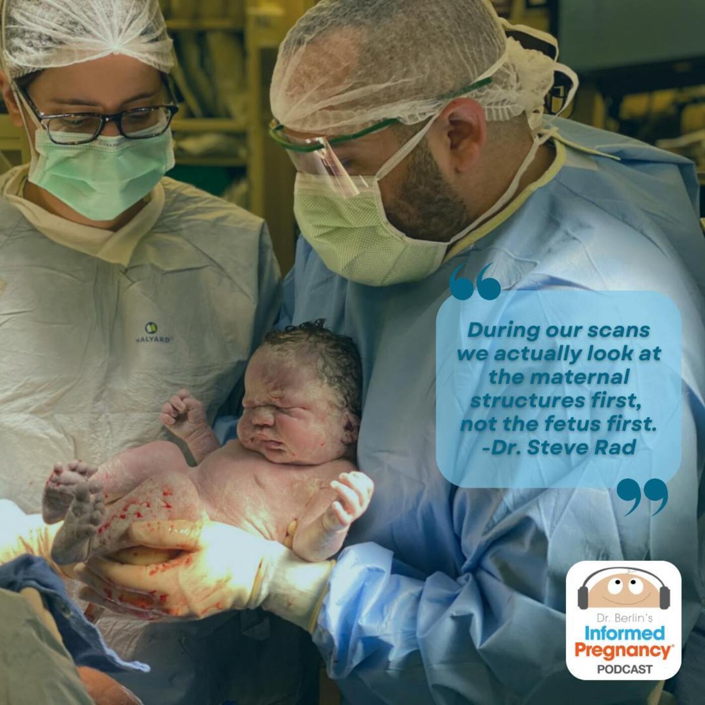
Dr. Steven Rad [19:49] Yeah. So we depend on the baby's position. We either start from the head down or from the feet up, we always go in order. And so you need to make sure you don't miss any And we memorize our protocols in the fetal anatomy by heart, but by going in order. So it's a safety guard. Anyway, so you first start out of the first thing you look at is, we actually look at the maternal structures first, not the fetus first. So I always look, make sure there's a heartbeat and a fetus and then we continue. Then I look at the mom's cervix, and I want to check, make sure the cervix looks long and closed. Then look at abdominally, we do do a vaginal ultrasound at the end.
[20:29] Some centers will start with a vaginal ultrasound and look at your cervix length first. Some places do the vaginal at the end, and some places will just look at your cervix abdominally. That's usually typically the first place at every…
Dr. Elliot Berlin, DC [20:42] What are you looking for at the cervix at the end?
Dr. Steven Rad [20:45] We want to make sure that the cervix is long, so there's no signs of cervical shortening or early labor. Okay. Next, this protocol should be typically universal when it comes to fetal specialists. So next thing you would look at is the placenta location. If it's anterior, posterior, if it's low lying or there's a placenta previa. And then at the placenta, you want to look at where the umbilical cord is coming out of the placenta, what we call the umbilical cord placental site insertion site. Typically it comes out of the middle of the placenta. If it comes towards the side, you would call it eccentric. If it comes off closer to the edge, it's called the marginal cord insertion.
[21:29] If it's not even attached directly to the placenta, it's called the filamentous cord insertion.
Dr. Elliot Berlin, DC [21:34] Filamentous?
Dr. Steven Rad [21:35] Yes.
Dr. Elliot Berlin, DC [21:36] Okay. This all sounds helpful and informative. Is there a practical difference whether the placenta is anterior in the front or posterior in the back?
Dr. Steven Rad [21:47] There is no difference for us. We care mostly if it's hovering the cervix or not. The difference is sometimes women whose placenta is anterior, they feel the fetal movements a little bit later in the pregnancy. Because as the baby, sorry, as the fetus grows, the placenta of its anterior, the top wall serves a little bit of a cushion, kind of like a pillow. Maybe it's kicking or punching into this placenta.
Dr. Elliot Berlin, DC [22:13] But no clinical difference.
Dr. Steven Rad [22:15] No, there's no clinical difference. There can be. Not necessarily. There can just be, from a patient perspective, a little delay in feeling fetal movements with an anterior placenta. It's not always the case. But there's no clinical difference for us. Unless you're doing a C-section. The only clinical difference is the time of delivery. You want to know where the placenta locations. For example, if you're doing a C-section, so if it's anterior and you want to know if you're going through it to get to the baby or if it's posterior, just for delivering and surgical purposes. But otherwise, it doesn't really matter.
Dr. Elliot Berlin, DC [22:45] So just collecting a data point.
Dr. Steven Rad [22:47] Yes, it helps though, the delivering the obstetrician helps if someone's scanning in the future where to look.
Dr. Elliot Berlin, DC [22:53] Okay, right. So for concern issues, you're looking to see if it's too low in covering the cervix placenta previa and how the cord is inserted to see if there's a problem there. And also other placenta anomalies?
Dr. Steven Rad [23:10] Sure, you can have placenta that's too thick or there's a placenta mass. There's placenta accreta in patients who have particularly previous C-sections or they have a placenta previa where that's where the placenta gets too stuck to the uterus and then bleeding at birth. So you want to look for signs of placenta accreta in specific clinical scenarios or if it looks abnormal. Placenta can be calcified in the placenta for the first or later under the pregnancy. I do want to point out one thing, since you brought that up, is the size of the placenta. The size of the placenta, not everybody looks at our measures. In our center, we take care of a lot of women with previous pregnancy loss or previous stillbirths.
[23:59] And one common cause of stillbirths was a small placenta that wasn't noticed. So our clinic is actually a certified rainbow clinic. This is the second in the U.S. In a small group internationally, we take care of women with previous stillbirths and pregnancy loss with a closer monitoring, a very strict protocol. And one of that part of that is to check the placental size and placental volume by a method that was developed by Professor Kleinman at Yale University, who personally trained me and came out to our site and we made a training video to try to train other providers. And there's a whole app by gestational age. So we look at the placenta volume as well and measure the size of the placenta.
[24:46] And patients with a smaller placenta have higher risk of fetal growth restriction, pregnancy complications, and stillbirths. So that's something that we look at closely.
Dr. Elliot Berlin, DC [24:55] OK, so we have the cervix and the placenta. the center and then what happens next?
Dr. Steven Rad [25:02] Yeah, in the bilibulcord, okay. In the bilibulcord and then next, we look at the next maternal structures, we look at the ovaries or at Nexa, on the sides to see if there's any cysts or like any cysts in the ovaries during uterine fibroids that we need to monitor during the pregnancy. So then those are the basic maternal structures that are looked at. Okay, then we move on to the fetus. Part of the fetus, we look at the amniotic fluid volume at the second trimester. You can look at it qualitatively, like subjectively, and we all know as we do ultrasound what's the normal amount of fluid or you can quantitatively measure the amount of amniotic fluid,
[25:46] what we call the single deepest vertical pocket.
Dr. Elliot Berlin, DC [25:49] Is the range of normal change from month to month?
Dr. Steven Rad [25:54] No, it's always two, we always want it to be between two and eight.
Dr. Elliot Berlin, DC [25:57] So there's both a quantitative and a subjective or qualitative. So you always want the deepest pocket to be between two and eight?
Dr. Steven Rad [26:06] Correct.
Dr. Elliot Berlin, DC [26:06] And then what about the AFI, the other way?
Dr. Steven Rad [26:10] You could do an AFI at 20 weeks, but typically to do an AFI, uterus is divided into four quadrants. Uterus isn't that big at this point. You could do one in theory, but the more proper thing to do is to just measure the deepest pocket. There has been studies showing that there's no difference in outcomes or significance between the deepest pocket and full AFI. So even before the studies never really measured an AFI at 20 weeks, some fetal centers or specialists may do that and you can still do that. Still the range is the same five to 25.
Dr. Elliot Berlin, DC [26:46] Okay, so that is the fluid.
Dr. Steven Rad [26:50] Yes, then I typically look to see that there's a three vessel umbilical cord. So there should be three vessels in an umbilical cord.
Dr. Elliot Berlin, DC [26:59] One vein and two arteries?
Dr. Steven Rad [27:01] Correct. And you can either look at that by looking at a piece of the umbilical cord and at a cross section of it, and you can actually see the three vessels, or you can look at the blood vessels around the bladder, the two arteries around the bladder to be present. And then you know that there's not what we call a single umbilical artery situation. So there's two vessels in the umbilical cord, one vein, one artery, or what's it called? So either two vessel cord, or another name for it is a single umbilical artery. So there's two ways to look for that. So we wanted the normal is three vessel umbilical cord. Then I personally, some folks might do later
[27:40] when they look at the fetal heart, but I check right away to see what's called the fetal sinus and make sure that I look at the position of the baby, or sorry, fetus, so see if it's like head down or if it's bridge or it's laying sideways transverse, and where the spine is, and we look to see where the heart and stomach and where all the organs are, and we make sure that the heart and stomach are on the left side of the fetus and the female body. So it's called a normal sinus, so I make sure the organs are on the correct side of the body. So that's where, when I do it, that might be different in other people's protocol where they look at that when they're looking at baby's heart and chest.
[28:23] So from there, then you go head to toe, or you go feet up and you go in order. And if you can't see something, you move on and skip it and come back, but you stay in order. You do not move around. Classically, students or new OBGYN residents or stenographers or people who don't do ultrasound often are just randomly looking around for a leg or a face or a head. We don't do that. We start in order, always in order. Okay, so when I'm looking at the fetal brain, we're looking at the shape and size of the head, and we're looking internally at the basic structures and making sure the brain is, for example, is divided in half and making sure the correct foldings are there and present,
[29:08] the amount of fluid is normal. And I look at the cerebellum, which is the bottom part of the brain. We look at the neck fold again, or the nuchal fold, and make sure all of that is looking good. Then from the head and brain, we move down to the face. In the face, we're not just looking to make sure the baby looks cute. We're looking at the orbits or the eye sockets. We're looking at the lenses of the eye, looking at the nose and nostrils, looking at the nose and lips and the palate, making sure there's no cleft-liver palate, looking to the profile and making sure that there's no abnormalities in the forehead or the actual nose and lip, and that entire structure,
[29:51] making sure that it's symmetric. And then looking at the chin, and then we might get a cute 3D at that point. Then, then we'll move down to the arms and look at the upper arms are humorous and we look at the forearms and the radius and all and then I look at the hands and count the fingers. And then we move on to look at the next so we make sure you notice, there's all the fingers and two hands and two arms. We move down and look at the baby's neck, make sure there's no masses or abnormalities in the baby's neck and the shape or position. Then we'll move down to the fetal chest and look at the chest appearance and size and in the chest we look at the heart.
[30:35] And like I said earlier we double check the side is or the position of things and make sure the hearts on the left side of the body. And the heart has four chambers and it has outflow tracks so a lot of connections between the heart and the valve. So we make sure all the heart is pumping well, it's structure and function, it's position, it's the heart rhythm, all that looks beautiful. Make sure the valves are looking good, making sure the arteries and veins coming off the heart and the structure large ones. For example, they go to the lungs or the aorta that takes blood to the upper body and to the lower body from the heart and the veins that bring blood back to the heart we look at all that.
[31:18] Make sure the blood vessels are crossing over each other correctly. There's a lot of detail in the fetal heart and such that you can be even seen by pediatric cardiologists and get a full fetal echocardiogram either by pediatric cardiologists or your maternal fetal medicine specialist may be trained in formal fetal echo. The heart is a complicated structure we look at the lungs around the heart as well. When we look at the diaphragm, which is a muscle that separates the chest from the abdominal cavity and is a muscle that helps you breathe and works on its own, goes up and down. So everyone should know the diaphragm is, make sure that diaphragm is present and that the organs in the stomach are separate from the organs in the chest there is an abnormality called diaphragmatic hernia where there's a hole in the diaphragm and some of the
[32:11] organs from the belly like your liver and stomach and end up in your chest. Then we move on to the abdomen and we measure the size, by the way the head we measure the size of the head I didn't mention that arms we measure the bones, measure the bones and arms and as we go down to the abdomen we measure the fetal abdominal circumference, which I think we've done a podcast on the fetal growth restriction together but so we measure the size of the fetal abdomen. In the abdomen there's organs, the main organs we look at are the stomach, liver, gallbladder, make sure there's two kidneys we look at for the bladder, make sure there's no dilated intestines or any abdominal masses or nothing's enlarged or blocked from these organs.
[32:57] And then around the bladder we talked about how you tell the three vessel umbilical cord. If you're looking at the two umbilical arteries which pass over the bladder before they go into the belly button which by the way we look at the umbilical cord insertion in the abdomen is what we talked about the abdominal cord insertion the placenta we also make sure that the umbilical cord is coming out. The abdominal wall is closed there's abnormalities where there can be an opening in the abdominal wall umbilical cord itself and the organs of the abdomen are just sticking out or spilling out. Let me make sure that the abdominal wall is completely closed and we look at the umbilical cord but that that's your belly button but it's all closed up and looks nice.
[33:42] We go down a little bit lower we'll go to the pelvis and we'll see the feel sex which will let you know if you want to know or hide it. And then we look at the legs so we look at the thigh bones and measure those we look at the leg bones and can measure those and we look at the feet and toes. And then lastly is the spine. You want to look at the spine appearance shapes.
Dr. Elliot Berlin, DC [34:06] My favorite part.
Dr. Steven Rad [34:07] And position. Yeah, and you want to make sure that there's a complete spine from top to bottom all the way from the neck down to the spine tip on the everything the spine is closed up. And one of the most common our eyes we look for a spina it's rare but one of the more everyone's worried about his spine a bit for that. Make sure there's no opening in the spine or the nerves are coming up and make sure that the entire skin line over the spine is closed.
Dr. Elliot Berlin, DC [34:34] That sounds like I could see why it would take an hour or more.
Dr. Steven Rad [34:37] There's a lot.
Dr. Elliot Berlin, DC [34:38] Yeah. So I don't know how the hell I don't know how you can do it in five.
Dr. Steven Rad [34:42] But there are some like you said maybe they just take a lot of pictures and do the analysis later. So, yeah, a lot of them do that and they're just a lot of fetal specialist if you've been doing this for like 3040 years your eyes are trained and you can do it really fast. It also depends on how you're saving it if you're saving each picture versus it's really. what I call like a tour of the human body from head to toe. And we look at it very carefully and we look at each organ carefully and it takes a long time because we want to get most beautiful images and make sure that everything is really in and following the protocol carefully. And we double check things.
[35:18] So we see things at least twice. And then if I'm not in the room, I just have one sonographer, Brittany, who a lot of patients know or might be listening know and love, but if she does it and I'm not there, I'll have her scan head to toe live for myself to see. And we'll double check everything but not with saving everything but she'll show me everything if I hadn't done the scan myself.
Dr. Elliot Berlin, DC [35:39] Okay, let's take one more break and we come back. I'm gonna ask you a few questions that people ask me and I'm not the right person to ask, but you are. We'll find out when we come back. Welcome back to the Informed Pregnancy podcast. We're finishing up our discussion of the anatomy scan with Dr. Steven Rad. You've already given us so much information, doc. Here's a couple of questions that people ask. Is there anything someone needs to do before the scan to get ready like sometimes for these clinical things? You want them to have a full blood or an empty blood or eat or don't eat?
Dr. Steven Rad [36:25] No, there is nothing you need to necessarily do. I would make sure you come having eaten because it can be long, bring maybe some water with you or a snack if you like. I think just knowing where you're going, how long they take is most, just kind of being prepared in terms of that because the hardest thing is this mom's schedule right before the other child needs to be picked up or right before bedtime or they need to be at another appointment. It can be pretty stressful to them. So we recommend that you plan accordingly depending on what you're quoted at the fetal center how long the scan will be. Like that's the biggest preparation you can do.
[37:09] So you're not in a rush or the person scanning you is not in rush or you're not angry. We don't want anyone to get angry or disappointed or whatever. So I think just planning on the time is the most important thing. In terms of empty bladder, full bladder, we have everyone empty their bladder before. We do their vaginal and the uterus is so long that we want them to be comfortable. And if we need their bladder to be full, it fills pretty fast. Oftentimes people need to even take a break in the middle. So we just have them empty their bladder from the beginning. So I think most centers, back in the, I think all the times they would tell you like,
[37:44] go drink a bunch of water before you come. And maybe some centers still have those rules but pregnant women's bladder fills pretty quickly.
Dr. Elliot Berlin, DC [37:50] What's the benefit of a full bladder for ultrasound?
Dr. Steven Rad [37:53] So if the bladder is full, you can see the cervix or placenta location or certain structures a little bit better, a full bladder. Sometimes it affects the position of the baby. So if the bladder is empty, there's a lot more room for the baby than when the bladder is full, then maybe the baby was more likely to stay in its position so you have more time to look at the spine, for example. But if you can only see the spine, you can't see the face or heart because the baby is looking down and showing its spine up top. We have to have you go empty your bladder and hope the baby slips. So really it doesn't matter for us. We start whatever, I mean, we have everyone empty their bladder
[38:33] and whatever we see, we see and we move forward. So like I said, we have an order that we go in but if the baby's position is not ideal, for example, to sit in that exact order, we come back to that structure or that organ system.
Dr. Elliot Berlin, DC [38:45] One more question for you, which is it seems like some people are nervous about doing ultrasound. It's not radiation. Is there any concern about doing ultrasounds or doing too many ultrasounds? Or would you tell somebody who's nervous about doing it and would prefer to skip?
Dr. Steven Rad [39:06] So Dr. Berlin, yeah, there's some very old studies that may have suggested ultrasound or too much ultrasound can be unsafe but there is no radiation, like you said. So our current knowledge, there's no adverse effects of getting ultrasound and the systems now are regulated and monitored for fetal safety and essentially if you're a certified center, you're one of the parts of certification is to show that you have the correct settings on your machine. And in terms of the number of ultrasounds, again, there's some like studies that showed that ultrasound can heat structure. But it would take many hours of a fetal ultrasound machine that's regulated
[39:50] to cause any, if ever, any tissue heating but it would have to be on there for many hours or months at the doctor's office doing that. I'm out of here, that's all. ultrasound, so the fetal ultrasounds rest assured to our knowledge are safe, there's no danger in them and are regulated and that's different than some of the therapeutic ultrasound procedures you might hear, so there are some procedures for whatever the conditions where they use ultrasound are very high energy and that's different than what you're getting in the fetal ultrasound, and lastly we don't recommend, it's not recommended to do ultrasound by the American College of Ob-Gyn, they don't, of course a lot of
[40:33] people do get these like 3D ultrasounds, there's even ultrasound centers, you just go just for 3D pictures, again they're usually really short visits, they give you like four or five pictures and you go, and technically they're safe, but it's not recommended to do ultrasound for fun for example, but of course many people want to do that, but even if you do their short visits it should be safe to be honest.
Dr. Elliot Berlin, DC [40:56] Dr. Rad, thank you so much for another great episode and lots of informative data about what to expect during an anatomy scan, when it's done, what it'll feel like, approximately how long it takes and what are the things that we're looking for, as always I appreciate your expertise and spending your time with us,
Dr. Steven Rad [41:17] where can we find you online? First of all thank you for having me, thank you for our listeners, I enjoyed that because ultrasound is one of my passions and what I do every day, all day, at least six days a week. You can find me online, the best place is on our Instagram at Dr. Steve Rad, and our office is in West Hollywood Beverly Hills at the Cedars-Sinai Medical Office Towers. Amazing, and I'd hope
Dr. Elliot Berlin, DC [41:45] thanks for listening to the Informed Pregnancy podcast. For more pregnancy and parenting information, visit us online at InformedPregnancy.com
Want more pregnancy + parenting? Start your FREE TRIAL of Informed Pregnancy+ and get access to all our curated pregnancy and parenting content — original series, iconic birth films like The Business of Being Born, prenatal workouts and more.